Tag Archives: ROM

post mastectomy physical therapy

Post Mastectomy Physical Therapy

Post Mastectomy physical therapy Exercises

Post Mastectomy Physical Therapy

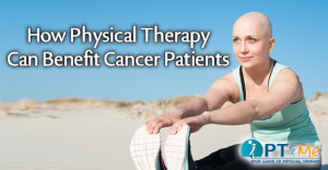
The word cancer is a scary one. Even though we all hope that it never becomes part of our lifetime of trials, more often than not, we know someone that has had, or is currently dealing with cancer. It is a testament to the medical community that so many women are able to wear the pink ribbon as a sign of triumph and pride, but we still mourn with those that wear it as a sign of remembrance and loss. More than once, while talking with women that have begun treatment for breast cancer, the topic of whether or not to have a mastectomy has come up. It’s not a decision taken lightly, often one with multiple concerns about what happens after surgery. Will the cancer be gone for good? Will it hurt? How long will it take to recover? A physical therapy post-mastectomy program can help address these issues.

Physical Therapy can’t answer all of those questions, but one thing a physical therapy post-mastectomy program can do is aid in the overall recovery process by focusing on regaining strength and increasing the range of motion in your shoulder and arm. Early intervention by a physical therapist can help women regain full function following mastectomy surgery, regardless of whether or not a woman has had reconstruction. Rehabilitation is always tailored to each patient’s specific needs. Not every patient experiences the same recovery, and as such physical therapists are prepared to help patients experiencing a multitude of symptoms – some have been highlighted below.

Size, location, and the type of mastectomy are important considerations when choosing a type of treatment. Exercises to maintain shoulder range of motion and arm mobility may be prescribed as early as 24 hours after surgery.  These exercises are important in restoring strength and promoting good circulation. As rehabilitation progresses these exercises may be modified to meet new goals.

Physical Therapy after Surgery

After mastectomy surgery, patients may experience tightness around the surgical site. This is caused by scar tissue formation. The result can be very dense tissue under the incision, which is painful and can restrict the range of motion.  The restricted range of motion puts a woman at risk for a painful condition known as frozen shoulder. Early treatment by a physical therapist can help reduce the pain and help regain functional range of motion and strength.

Numbness and/or nerve sensitivity at the surgical site can develop post-mastectomy. Manual therapy can help restore sensation and relieve nerve pain. In severe cases, a chronic condition known as post-mastectomy pain syndrome may develop.  This is caused by scar tissue impinging on nerves. Physical therapy can be very effective at releasing scar tissue and reducing nerve-related pain.

Axillary node dissection can lead to a condition known as cording or axillary web syndrome.  Cording presents as a moderate to painful tightening, which appears as “cords” emanating from the armpit and extending down the arm. Cording significantly restricts the range of motion and arm function. Manual therapy and therapeutic stretching help to resolve this condition quickly.

Radiation treatment after mastectomy surgery can exacerbate posture and range of motion problems, causing fibrosis and skin tightness. Manual therapy can remediate these issues and may prevent them from ever becoming a problem.

The Benefits of Exercise and Physical Therapy post-mastectomy treatment programs can differ greatly as seen above, but there are a few benefits that all patients can benefit from:

  • Improved shoulder range of motion
  • Improved shoulder strength
  • Improved functional mobility
  • Improved posture
  • Decreased pain at the surgical site
  • Decreased edema on the affected side
  • Improved sensation at the surgical site

Meeting with a physical therapist before surgery can help you feel more at ease and more confident in your overall recovery goals. It’s never too early to ask questions! To find a physical therapy clinic near you click here.

For more information on cancer-related physical therapy programs click here:


physical therapy near me
Physical Therapy for Golfer's Elbow

Golfer’s Elbow

Physical Therapy for Golfer's Elbow (Medial Epicondylitis Pain)

Golfer’s Elbow

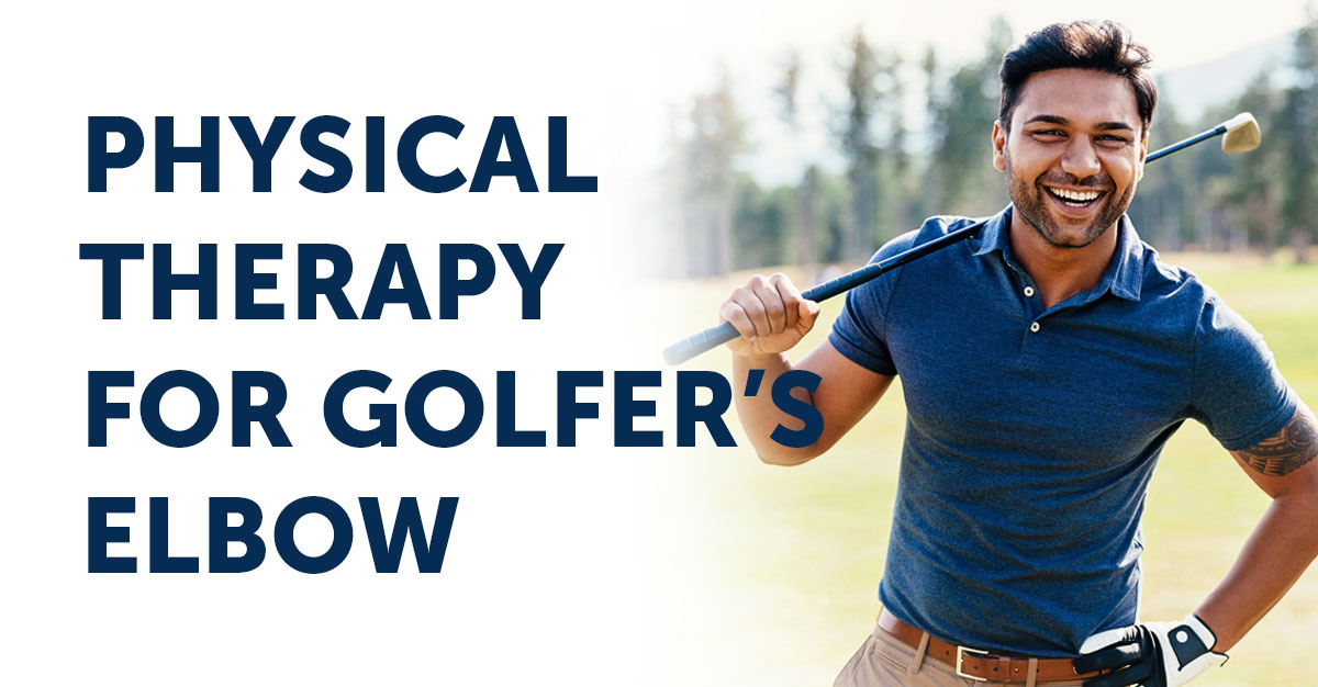
Golfer’s Elbow, medically known as medial epicondylitis,  is a painful condition where the tendons that attach to the inside of the elbow become inflamed due to repetitive use of the hand, wrist, forearm, and elbow. Golfer’s elbow often occurs with repetitive activities such as swinging a golf club or tennis racket, work or leisure activities requiring twisting and gripping such as shoveling, gardening, and swinging a hammer. Golfer’s elbow can also appear in other sports-related activities such as throwing and swimming. Golfer’s Elbow (Medial epicondylitis) is most commonly seen in men over the age of 35 but can be seen in any population. If these symptoms sound familiar, then going to physical therapy for golfer’s elbow may be just what you need.

Physical Therapy Appointment

What is causing your elbow pain?

Golfer’s Elbow (Medial epicondylitis) affects the group of muscles that are responsible for bending the wrist, fingers, and thumb and that rotate the wrist and forearm. The tendons that connect those muscles to the medial epicondyle (bump on inside of elbow). Tendons are made up of collagen fibers that are lined up next to each other. The repetitive forces pull on those tendons creating pain and tenderness described as Golfer’s Elbow. Without treatment, those tendons can eventually pull away from the bone. Acute injuries to your elbow can create an inflammatory response which can cause redness, warmth, and stiffness in your elbow.

Golfer’s Elbow (Medial epicondylitis) is most often caused by an abnormal arrangement of collagen fibers. This condition is called tendinosis. During tendinosis, the body doesn’t create inflammatory cells as it does during an acute injury. Instead, fibroblasts are created which help make up scar tissue to fill in the spaces between the collagen fibers. This increase in scar tissue can lead to increased pain and weakness in the tissues. Physical and hand therapy is the most common nonsurgical treatment for Golfer’s Elbow (medial epicondylitis). Your therapist will perform an evaluation where he/she will ask you several questions about your condition, pain level, and other symptoms you may be experiencing. He/she will perform motion and strength testing on your entire upper extremity. Your therapist will also palpate your arm to determine which tendon(s) may be inflamed. He/she will use special tests designed to deferentially diagnose your condition from others that may have similar presentations to Golfer’s Elbow, such as Cubital Tunnel Syndrome.

golf ball on tee

What to Expect from Physical Therapy for Golfer’s Elbow

  • Pain Management: this can include Mechanical Diagnosis & Therapy, ice, ice massage, moist heat, electrical stimulation, and ultrasound.
  • Range-of-Motion Exercises: stretches and mobility exercises to help maintain proper movement in your elbow, forearm, wrist, and hand.
  • Strengthening Exercises: progressive resistive exercises to help build strength in your arm, elbow, forearm, wrist, and hand. These can include weights, medicine balls, and/or resistance bands. This will also include your Home Exercise Program.
  • Manual Therapy: used to ensure full, pain-free movement is achieved and can include joint mobilizations, manual muscle stretches, and soft tissue massage.
  • Neuromuscular Re-education (Functional Training): used to help you return to your prior level of function for both home and work activities. This will include retraining proper movement patterns with necessary modifications based on the current level of function and patient limitations.
  • Patient Education: used to help retrain patients on proper postural control during everyday activities including dressing, self-care, work, and sports activities. This can include helping return a patient to their specific sport, such as making adjustments to their golf swing or throwing technique.

Once you’ve completed physical therapy for Golfer’s Elbow you’ll want to do everything you can to prevent this from reoccurring. This can occur by maintaining proper awareness of your risk for injury during your daily movements. Key things to keep in mind:

1. Maintain proper form during all repetitive movements both at work and at home.
2. Continue your Home Exercise Program in order to maintain proper strength in your shoulder, elbow, forearm, wrist, and hand.
3. Use proper posture and body mechanics with lifting or carrying to avoid any undue stress on your joints and tendons.

This information was written by Plymouth Physical Therapy Specialists, an outpatient physical and hand therapy group with fourteen locations in the surrounding Plymouth, Michigan area. At Plymouth Physical Therapy Specialists, they are committed to using evidence-based treatments in their practice. This means that their therapists utilize the most current and clinically relevant treatments in their approach to rehabilitation. For more information click here.

physical therapy near me

shin splints

7 Ways Physical Therapists Treat Shin Splints

Ways physical therapists treat shin splints.

Here are 7 ways a physical therapist can help treat pain and symptoms associated with shin splints:

Pain Reduction: The RICE principle is the first step to recovery (rest, ice, compression, and elevation). Manual therapy and Kinesiotaping may also speed up recovery and reduce swelling.

Gait and Footwear Analysis: An analysis of how a person walks and runs is an important part of treatment. The wrong mechanism of walking can transmit a great deal of force through the shin to the knee and hip. In such situations, physical therapists will correct gait patterns and recommend footwear with shock-absorbing capacity.

Muscle Stretches and Strengthening: The tibial and peroneal muscles are attached to the shin and must be stretched adequately before any form of exercise. Physical therapy includes various stretches of the foot that will help stretch and warm up these muscles. Strengthening damaged muscles can also help.

 Activity Modification:  Physical therapists may suggest alternative activities to minimize stress on the shinbones. These can include swimming and cycling.

Increase Range of Motion (ROM): Exercises for the hip, knee, ankle, and foot improve blood circulation, reduce inflammation and relieve pain. A home exercise program may also be implemented.

Arch Support:  The absence or collapse of a normal foot arch can lead to shin splints. Physical therapists will recommend appropriate orthotics that can be custom-made for the patient and provide the proper amount of arch support.

Return to Sport: If you are an athlete, your therapist may tailor exercises that are specific to strengthening the areas needed to perform your sport. Modified use of your muscles may also be discussed and implemented. Return to your sport may be gradual to prevent re-injury.

To learn more about shin splints please visit our PTandMe injury center on this website by clicking here.

Physical Therapy Appointment

total knee replacement

Physical Therapy Following a Total Knee Replacement

Physical therapy following a total knee replacement

There are over 600,000 total knee replacements done each year in the U.S. As the workforce ages and as obesity levels continue to rise, this number is expected to increase.

Candidates for a knee replacement include patients experiencing difficulty doing simple daily activities, including walking or going down steps, and where conventional treatments are no longer helping. Common causes that lead to a replacement include pain with simple ADL’s (activities of daily living), pain at rest, chronic swelling, inflammation not improving with medication, knee deformity, knee stiffness, or trauma to the joint. When you are scheduled for a total knee replacement, ask to see your physical therapist for a pre¬surgical exercise and stretching program. This will help your knee recover its range of motion and strength.

Surgery consists of resurfacing the knee’s damaged and worn joint surfaces with artificial parts made of metal or plastic. Most patients are in the hospital for an average of 3¬5 days. And more than 90 percent of people report significant decrease in pain and improvement in ability to perform ADL’s within one month after surgery. Currently, many knee replacements are lasting 20 years or more with appropriate activity modification. One of the most important factors in success after a total knee replacement is follow up with physical therapy and a lifelong exercise program. Physical therapy typically starts during the hospital stay with the goals being to get the individual up and walking decreasing swelling in the knee and increasing knee ROM (range of motion). Upon hospital discharge, patients either go home and receive home physical therapy for one to two weeks, or to an extended care facility to continue their therapy.

Typically, patients need outpatient physical therapy after therapy at home, which usually begins two to four weeks after surgery. Outpatient physical therapy is recommended for three times a week for up to three months. Therapy goals initially are to reduce swelling and pain, and improve knee range of motion in both directions. To achieve these goals, treatment may consist of soft tissue massage to increase circulation and decrease swelling, stretching to improve flexibility, patellar mobilization, range of motion exercises, and modalities such as ice and electrical stimulation to help decrease pain and swelling. Physical therapy will progress to lower extremity strengthening exercises, balance activities, and fine tuning the gait pattern. Various activities and techniques will be used to improve strength, balance, and gait. As patients reach the end of their course of physical therapy, a personalized home exercise program is developed with the focus on independence with all ADL’s and returning to activities such as walking, swimming, dancing, golf, and biking. It is important for patients to stay active and maintain strength, flexibility, and endurance.