There are several types of arthritis, which can be defined as an inflammation, degeneration, or infection of a joint. Some types of arthritis just affect a few joints and are called oligoarthritis. Others affect many joints in the body and are called polyarthritis. Arthritis can be symmetric, meaning the pain and inflammation is roughly the same on both sides of the body. It can also be asymmetric, affecting just about any joint in the body at any given point in time. This article will talk about several of the major types of arthritis, including osteoarthritis, rheumatoid arthritis, septic (infectious) arthritis, and crystal arthritis (which includes gout).
OSTEOARTHRITIS (OA)
Osteoarthritis (OA) is slowly-developing form of arthritis that can affect nearly every joint and that can affect individuals as they age. Joints that take a lot of pressure or that have repetitive motion are at the greatest risk for OA. The disease can be relatively asymptomatic or severely debilitating, affecting the patient’s ability to participate in activities of daily living.
The primary problems with OA are joint pain and stiffness. Symptoms can affect just one joint or several joints. The pain is worse with movement and relieved by rest. There are three stages of pain. The first is stage 1 (predictable, sharp pain with movement). The second is stage 2 (more constant pain that affects daily activities). The third is stage 3 (constant dull/aching pain with unpredictable sharp flares). The pain is usually worse in the late afternoon and evening but can also be worse after awakening. It may interfere with sleep.
OA can be localized or generalized and can affect one single joint or multiple joints at a time. The joints that are particularly a problem for OA include the knees, hips, interphalangeal (hand) joints, facet joints of the neck and back, first great toe joint, and thumb joints. The other joints are less commonly affected, particularly if they are not moveable joints.
Patients with generalized arthritis usually have the distal interphalangeal joints of the fingers involved, the base of the thumbs, the first great toe joint, the spinal facet joints, knees, and hips. Gradually, more joints become involved and the patient becomes more debilitated. The main clinical marker for OA is Heberden’s nodes, which are hard lumps at the distal finger joints.
Diagnosis of OA
Imaging can help diagnose OA. The best test for osteoarthritis is the conventional x-ray. It allows for detection of the usual features of OA (including osteophyte formation, cysts on the bone, and joint space narrowing). An x-ray, however, isn’t very sensitive and doesn’t often correlate with symptoms.
As to specific joints seen on x-ray, the hands are usually bilaterally and symmetrically involved. The distal interphalangeal joints at the tip of the fingers are characteristically affected. Other joints affected are the other finger joints and the base of the thumb. There is a lot of aching and stiffness of the joints. The patient with Heberden’s nodes has nodal OA. Most individuals with nodal OA are female with a family history of the disease.
Erosive osteoarthritis is a rare but aggressive type of hand OA. There is subacute or insidious pain with soft tissue swelling and numbness of multiple finger joints. There is tenderness, redness, and soft-tissue swelling of the hands that is not seen in ordinary OA. Erosive OA is not connected to generalized OA as it affects mostly the finger joints and spares the thumb and metacarpal phalangeal joints. Erosive OA has a worsened outcome than regular OA and can be seen on x-ray showing joint erosions (wearing down) in up to 8% of patients.
The knee is a common site of OA and the most common cause of lower-limb disability in older adults. It is usually bilateral to some extent. The patellofemoral joint or the medial tibial femoral joint is most affected. Pain from patellofemoral joint OA is made worse by prolonged sitting, standing up from a low chair, and climbing stairs or inclines (coming down often being more painful than going up).
Osteoarthritis of the hip can be seen as increased pain, stiffness, aching sensation, and restricted movement of the hip joint. Pain secondary to hip arthritis is felt in the anterior groin but may involve the upper thigh and buttocks. It commonly radiates down the leg with thigh pain and knee pain common complaints. The pain is made worse by rising from a seated position and during the initial phases of walking.
Facet joint arthritis generally goes along with intervertebral disc degeneration—a term called “spondylosis”. The pain is localized primarily to the lumbar or cervical spine and, in the low back, the pain radiates to the groin, buttock, or thighs, ending at the knees. It is worse in the morning and when active with rotation or bending motions (or with neck rotation and lateral flexion in the neck).
OA can be differentiated from other diseases by clinical history and physical examination. Rarely are things like lab tests and x-ray recommended. The clinical findings are of persistent pain in overused joints, age older than 45 years, and stiffness of less than 30 minutes in the morning. Imaging and lab tests can be done if the person doesn’t meet the obvious clinical features (by history or physical examination). Constitutional symptoms (like weight loss and fatigue) or signs of inflammation of the joint point away from OA and need further evaluation.
Risk Factors for OA
OA has been found to be a complex interaction of many factors, including genetics, mechanical forces, joint integrity, and certain biochemical processes. Genetics is probably the rarest interaction, while things like occupation, aging, trauma, and repetitive movement play a stronger role. These are stronger for the hand and knee and less strong for OA of the hip. Common risk factors include age, being female, being obese, having no osteoporosis, certain occupations, playing certain sports, having an injury, muscle weakness, and proprioceptive deficits. Less common risk factors include genetics, having acromegaly, and having CPPD disease. Advancing age is the strongest risk factor. It occurs in less than 0.1 percent of those under 34 years but is present in more than 80 percent in those older than 55 years.
Previous injury seems to increase the risk of osteoarthritis of the knee and having congenital hip dysplasia enhances the risk of hip arthritis. Long-distance runners have an increased risk of knee injury and knee osteoarthritis. Having an injury during this sport will increase the risk of osteoarthritis of the knee. Knee meniscus injuries are common in OA of the knee. Having an amputation of one leg increases the pressure to the other leg and increases the chances of OA of the unaffected leg. Genetics play a small role in getting osteoarthritis. There is a genetic influence in getting osteoarthritis of the hands and knees.
Treatment of OA
The goals of the treatment of osteoarthritis are to decrease pain, improve function, and modify the process of joint damage. This depends on changing modifiable risk factors as there are no disease-modifying OA drugs. Usually a combination of treatments is recommended. Things like hyaluronic acid injections in the knee are not recommended because they do not work any better than placebo.
The mainstay of treatment for OA is nonpharmacologic interventions. These include weight management, orthotic devices, braces (if necessary). Exercise has been found to be as good as NSAID therapy with strengthening and aerobic exercises good choices. A loss of 10 percent of the body weight will decrease pain by 50 percent with knee arthritis and hip arthritis. Splints and knee braces are good for thumb and knee arthritis, respectively.
Second-line things for osteoarthritis include drugs, such as capsaicin, nonsteroidal anti-inflammatory drugs, duloxetine, and intra-articular corticosteroids. A combination of these can be tried. Duloxetine is also called Cymbalta, which is an SSRI antidepressant that works for arthritic and musculoskeletal pain disorders. If a few joints are affected, a topical NSAID is recommended, with oral NSAIDs used only if topical medications don’t work. Acetaminophen has a risky side effect profile and a negligible effect on OA pain, so it isn’t recommended. Opioids are not recommended as they don’t work well for OA and have a long-term dependence and abuse potential.
Surgery usually means total joint replacement—usually done for advanced hip and knee arthritis. Other surgical options include a partial meniscectomy or debridement of cartilage but these have no clinical benefit over placebo. Hip arthroscopy can be done but may not be beneficial in OA.
RHEUMATOID ARTHRITIS (RA)
RA is a symmetric, inflammatory, peripheral arthritis, affecting many joints. The untreated patient will have degeneration of the cartilage and deformities of the joints in a symmetrical way. The prompt recognition and treatment of the condition with DMARDs, which are disease-modifying antirheumatic drugs, will help manage but will not cure the disease. The presentation in the beginning is similar to other arthritis patients but, over time, there will be distinctive evidence of RA, with joint erosions, extraarticular manifestations, and rheumatoid nodules.
Clinical Findings in RA
The synovial joints are what are affected most in RA. The arthritis is usually symmetrical, leading to destruction of joints secondary to bony and cartilaginous erosion. It starts in the hands and feet and moves centrally so locomotion becomes difficult within 10-20 years after onset. The onset is gradual and involves many joints, although some people will have a single joint involved in the beginning. Systemic symptoms occur in about 33 percent of patients and include muscle aches, low-grade fever, depression, weight loss, and fatigue.
In “classic” RA, the patient has morning stiffness, joint pain, and swelling of joints. The MCP (metacarpophalangeal) joint and the PIP (proximal interphalangeal) joints of the hand are the main joints involved initially; however, a few patients can have thumb, wrist, or metatarsophalangeal (MTP) joint involvement. Eventually other synovial joints of both the upper and lower limbs eventually become affected. Morning stiffness is the most common feature of active RA. It tends to last longer than an hour in RA and less than an hour in people with other inflammatory diseases.
Physical signs and symptoms include joint pain and swelling of the small joints (primarily), plus the typical morning stiffness and decreased grip strength. The spine is usually not involved. There is progressive joint damage and deformities, with loss of physical impairment. Late findings of untreated disease include anemia, rheumatoid nodules, eye inflammation, blood vessel inflammation, neuropathy, and pericarditis.
The hands are typically involved at the MCP and PIP joints. Redness and thickening of the flexor tendons may be seen in the palm; nodules may be seen in these tendon sheaths, causing trigger finger and possible tendon rupture. In established RA, there may be an ulnar deviation of the MCP joints.
The second most common areas of involvement are the wrists. Loss of extension happens early on in the disease process and, later on, there is volar subluxation and radial drift of the wrist. The elbow may become fixed in the flexed position. Olecranon bursitis is very common. Shoulder involvement is a late finding, seen in just half of patients after 15 years.
Lower extremity involvement is usually with the forefoot and ankles. Hip involvement is a late finding. Knee involvement can lead to Baker’s cysts. The MTP joints of the feet are the primary joints in early disease with eventual lateral drift of the toes and plantar subluxation of the metatarsal heads. Heel pain will show itself and the ankle may be swollen. Knee swelling is also common and restriction of flexion can be seen. There will be weakness of the quadriceps muscles.
Lab and Imaging Studies in RA
Lab findings in RA include those things seen in the synovial fluid and blood, indicating that the disease is both local and systemic. Things that are seen include inflammatory joint fluid, anemia of chronic disease, and lab tests that are positive for rheumatoid factor (RF) and ACPA (anti-citrullinated peptide antibodies). About 80 percent of patients will be positive for RA and/or ACPA. About 25 percent will have a positive antinuclear antibody titer.
Plain films can tell a lot about the state or RA. There will be joint space narrowing and bony erosions—especially of the hands and feet. These erosions are cardinal findings in RA. MRI testing is more sensitive in detecting synovial inflammation. It is also more sensitive for bony erosions than plain films. Ultrasound is also sensitive for detecting joint inflammation. Doppler ultrasound is nearly as good as an MRI and is cheaper than the MRI examination.
Evaluation of Suspected RA
This disease is usually present in adults and the main finding will be inflammatory polyarthritis. The affected person will have joint pain and at least thirty minutes of stiffness in the morning. Peripheral joints tend to be prominently involved. Symptoms lasting less than six weeks might be a viral polyarthritis instead of RA. In such patients, an anti-cyclic citrullinated peptide (CCP) antibody titer, rheumatoid factor, and acute phase reactants can be done. It may take many visits to get a clear diagnosis.
The examination includes a thorough joint evaluation, expecting symmetric polyarthritis, limited ROM of the muscles, and some extraarticular findings, like rheumatoid nodules. The lab tests will often include an RF and anti-CCP antibodies as a positive result that will increase the chances of it being RA. In an initial evaluation, however, these will be positive only in 50 percent of patients with early disease.
Other tests that are done include the ANA titer (which can exclude lupus and other rheumatic diseases). The ANA titer, however, will be positive in a third of RA patients so follow-up testing, like the anti-dsDNA and the anti-Smith antibody test should be done as these are highly specific for lupus. The CBC is done to check for anemia of chronic disease, liver and kidney function tests are done, and a serum uric acid level is drawn.
Baseline plain x-ray will be done of the hands, feet, and wrists in order to document a baseline so as to monitor disease progression. Joint erosions may or may not be seen initially. There are other specific findings seen in other joint diseases that will point to other diagnoses as well. Arthrocentesis is done to exclude crystal disease like gout. Gram-staining, cell counts, crystal search, and cultures are done on the fluid. MRI and ultrasound are not routinely done but they are more sensitive tests and can be done in patients with normal plain x-rays.
Treatment of Rheumatoid Arthritis
The treatment of RA depends on controlling the synovitis in the joint and preventing injury to the joint. Treatment strategies have changed remarkably over the last twenty years with the institution of DMARD therapy earlier in the course of the disease process. The goals include early diagnosis, care by a rheumatologist, early use of DMARDs, and tight control having a goal of remission or significantly reduced activity. Now, NSAIDs and glucocorticoids are adjunctive therapies instead of primary therapies. DMARDs have become the primary therapy.
Making the diagnosis as early as possible is important because DMARD therapy works best if there isn’t any joint damage. Once diagnosed, the patient needs a rheumatology referral and follow-up care performed by a rheumatologist (as the disease outcome is better). These patients need comprehensive care that includes drug therapy, education, psychosocial interventions, physical and occupational therapy, nutrition counseling, screening for osteoporosis, and things like vaccines to prevent disease in their immunosuppressed state.
Therapies include NSAIDs and intraarticular steroid injections, biologic and nonbiologic DMARDs, and an oral janus kinase inhibitor. Conventional, nonbiologic DMARDs include hydroxychloroquine, sulfasalazine, methotrexate, and leflunomide. There are a number of biologic DMARD drugs, including TNF-alpha inhibitors (etanercept, infliximab, adalimumab, golimumab, and certolizumab pegol), anakinra (an IL-1 receptor antagonist drug), and tocilizumab (an IL-6 receptor antagonist drug).
DMARD therapy is started as soon as possible. With active RA, an NSAID and corticosteroid are used along with methotrexate (as a first line agent). Patients who can’t take methotrexate should have hydroxychloroquine, sulfasalazine, or leflunomide. Nonbiologic DMARDs can be taken with biologic DMARDs. NSAIDs and prednisone are used temporarily as adjunctive therapy.
RA will naturally have flareups that need management. DMARD therapy may need adjusting. Flareups of just one or a few joints can be treated with intraarticular glucocorticoid injections. Widespread flareups are treated with an increased glucocorticoid dose (oral or IM). IV methylprednisolone done three times daily can be effective in reducing a flareup. Increased doses of methotrexate can help as will increased doses of infliximab.
SEPTIC ARTHRITIS
Septic arthritis is an infection in the joint, usually caused by a bacterial organism; however, it can be caused by mycobacterial species or fungi. These types of infectious processes can result in severe joint destruction and later arthritis. Among adults presenting with an acutely painful joint, septic arthritis represents 8-27 percent of cases, depending on the location in the world. The average is about 10 percent of cases. Some patients will have gonococcal disease, while others will have prostheses that get infected.
Risk Factors for Septic Arthritis
About ten percent of cases of patients with an acutely painful joint have septic arthritis. Risk factors for the disease include age greater than 80 years, having a diagnosis of diabetes mellitus, having rheumatoid arthritis, having a prosthetic joint, having recent joint surgery, IV drug use, being an alcoholic, having a concurrent skin infection, and having a previous corticosteroid injection. Each of these risk factors is small but together they add up.
If a patient has bacteremia, they are more likely to have the bacteria harbor in an arthritic joint when compared to a normal joint. About 40 percent of septic arthritis patients had preexisting RA or OA in the joint affected by sepsis. RA predisposes the patient to septic arthritis to a greater degree than OA but less common joint problems, like gout, pseudogout, and Charcot joint disease can be predisposing factors. Being on immunosuppressive therapy for RA is a predisposing factor, making RA a slightly higher risk than the other joint diseases.
Most cases of septic arthritis come from a blood infection that spreads to the joint. Trauma, direct inoculation, extension of bone infection, or animal/human bite are other less common causes. Risk factors for bloodborne spread include IV drug use, the presence of indwelling catheters, immunocompromised states (like HIV), being a neonate, and being older.
When the infective organism is Staphylococcus aureus, enterococcus, or streptococcus, endocarditis should be suspected and IV drug use is the most common reason for this infective endocarditis. Most of these patients will have negative cultures of the blood because the bacteremia was transient and self-limited. It is unknown why only a small percentage of patients with bacteremia develop septic arthritis.
Bacterial arthritis can occur along with bacterial meningitis. The most common organism associated with both septic arthritis and bacterial meningitis is meningococcus. Rare cases of septic arthritis can stem from catheter insertion (such as hip arthritis and sternoclavicular arthritis). A ruptured colonic diverticulum can also cause septic hip arthritis by spreading through the tissues.
While many pathogens can cause nongonococcal bacterial septic arthritis, the most common bacterial species causing this in adults is MRSA (methicillin-resistant S. aureus). Less common organisms include S. pneumoniae, Enterococcus, Escherichia coli, and other gram-negative bacilli. Almost all cases are from a single organism (except for those caused by penetrating trauma to the joint space, polymicrobial bacteremia, or ruptured diverticulum causing a hip infection).
Clinical Signs and Symptoms of Septic Arthritis
These patients tend to present with a single inflamed, painful, and swollen joint. There is usually warmth and restricted movement. Crepitus would not be noticed and there are no nodules. Many patients are feverish but will not have chills or spiking fever. There may be evidence of other infections present that point toward the original source of the infection.
The knee is involved in more than half of all septic arthritis cases, with lesser joints being the hips, ankles, and wrists. Rarely, the pubic symphysis can become infected after pelvic surgery. About 20 percent of the time, the situation is oligoarticular or polyarticular, especially with those individuals having RA or another connective tissue disease (and in severely septic patients).
Diagnosis of Septic Arthritis
The definitive test is to identify the bacteria in the synovial fluid. There should be a joint aspiration before starting antibiotics with synovial fluid sent for Gram stain, culture, leukocyte count (and differential), and crystal analysis. The tap may need to be done under fluoroscopic, ultrasound, or CT guidance. The results of the fluid analysis would be as follows:
• There will be purulence with a WBC count of 50,000-150,000 cells per microliter (mostly neutrophils). The higher the WBC count, the greater is the chance of sepsis.
• The Gram-stain is positive in 30-50 percent of cases.
• The culture should be done and will be positive in most patients who haven’t recently been on antibiotics. Blood cultures are generally positive in 50 percent of cases so they should be done.
Treatment of Septic Arthritis
The two components of acute bacterial arthritis include antibiotics and drainage of the joint. The choice of antibiotic depends on the presentation, Gram stain, and probable organism. Gram-positive cocci deserve vancomycin as an empiric treatment as this might be MRSA. If it is found not to be MRSA but to be methicillin-susceptible S. aureus, then the agents of choice include cefazolin, nafcillin, or oxacillin. Second-line treatments for MRSA include daptomycin, linezolid, or clindamycin.
If the Gram stain shows gram-negative bacilli, the treatment is a third-generation cephalosporin, such as ceftriaxone, cefotaxime, and ceftazidime. IV drug users are at risk for Pseudomonas infections and should have a combination of ceftazidime or ciprofloxacin plus gentamicin (or another aminoglycoside). Immunocompetent patients with negative gram stain should be treated with vancomycin. Ultimately, the culture and sensitivities will help detect the correct antibiotic choice. Intraarticular antibiotics are not recommended.
GOUT
Gouty arthritis is a type of arthritic condition that causes extreme pain and swelling in the joints. It stems from having high uric acid levels in the bloodstream that form crystals of monosodium urate (MSU) in the joint space. The crystals are very irritating to the joint and will cause swelling and redness of the affected joint space. About a third of all patients with high uric acid levels will develop gouty arthritis. It isn’t clear why some people get gouty arthritis and why others will not get gouty arthritis.
It should be noted that the joints aren’t the only place where uric acid crystals can develop. They can precipitate in the kidneys (impairing kidney function) or in other parts of the urinary tract, causing pain and possible obstruction of the urinary system.
Gout is completely different from CPPD (calcium pyrophosphate dihydrate) deposition disease or “pseudogout”. The crystals are completely different and the etiologies are different. Some of the symptoms are the same, however, so it pays to have an arthrocentesis and crystal analysis in order to define what type of crystal disease a person has.
Risk Factors for Gout
Gout is an adult disease that usually is first manifest in men between the ages of 30-45 years or in women over the age of 55 years. There is no gender difference above 65 years. The prevalence is about 4 percent of all adults in the US. Risk factors for gout include hypertension, obesity, chronic renal insufficiency, fasting, and consuming alcohol on a regular basis. Overeating, especially those things with high fructose corn syrup, meats, and seafood, will increase the risk of gout. Diuretics will increase the uric acid level.
Flareups of gout in patients already known to have gout can include fasting, recently having surgery or an injury, drinking too much alcohol (especially wine), overeating, and taking certain medications.
Symptoms of Gout
Gouty arthritis flares or gout attacks involve the sudden onset of joint pain that is relatively severe and associated with tenderness, swelling, and redness of the joint. It usually affects one joint but can affect a few. The flareups are worse in the middle of the night and in the early morning hours. The inflammation reaches its peak at 12-24 hours and can improve within a few days or weeks. No one knows how the body resolves a gouty flare. The white blood cells increase in the joint space because of the crystals causing irritation of the lining of the joints.
Gouty Arthritis Phases
There are three basic phases of gout: the gout flare, intercritical gout, and tophaceous gout. In a gout flare, the big toe or knee becomes inflamed to begin with or multiple flareups can occur—accompanied by a fever. Some osteoarthritic patients will have flareups in the fingers instead of the toes. Intercritical gout is the time between flareups, which is generally less than two years, even with treatment.
Tophaceous gout is the type of gout where urate crystals build up around the joints, bursae, skin, bones, and cartilage. The buildups are called “tophi”. They may have bony erosion and joint damage called gouty arthropathy as a result. They usually aren’t painful but can inflame like joints, causing redness and tenderness. Tophaceous gout is rare with current treatment protocols except if a person cannot tolerate the medications, is taking cyclosporine for organ transplant, or is a woman past menopause on diuretic pills.
Diagnosis of Gout
Gout can mimic many other diseases. It is strongly suspected when a joint or few joints become acutely inflamed and then resolve within a few days, leaving behind no symptoms. The best way to confirm gout is to take a quantity of synovial fluid using an arthroscopy. The fluid can be evaluated under specialized light microscopy to see if the characteristic needle-shaped crystals can be seen. There will also be an excess of white blood cells in the synovial fluid. Crystals can be extracted from tophi as well. If arthroscopy cannot be done, the diagnosis is based on acute onset of symptoms, inflammation of one joint (usually the great toe), elevated uric acid levels, and complete resolution of symptoms between flareups.
Treatment of Gouty Flareups
The goal of treating gouty flareups is to decrease pain and inflammation. This is a short-term process that is based on the presence or absence of bleeding disorders, history of stomach ulcers, or history of kidney disease. If these are negative, NSAID drugs are the treatment of choice as well as glucocorticoid drugs. Corticosteroids can be given orally, injected into the joint or by injection. Commonly-used glucocorticoids include prednisolone, prednisone, and methylprednisolone. They can be used when NSAIDs or colchicine aren’t tolerated.
NSAIDs are okay if there are no bleeding problems (no warfarin), no stomach ulcers, and no kidney disease. Aspirin is not recommended because it can affect the uric acid level in the bloodstream. Colchicine is good with decreased kidney function and ulcer risk but can cause abdominal symptoms (like nausea, vomiting, diarrhea, and abdominal cramps). It is only taken orally.
There are preventative drugs that will decrease or reduce the number of gouty flareups. Colchicine is one drug that can do this and can be given in lower doses than is necessary to treat gouty arthritis flareups.
Some patients require long-term uric acid-lowering treatment. Lifestyle changes can help the situation. Medications can increase the uric acid excretion by the kidneys, decrease the production of uric acid, or can convert urate to allantoin (which is more easily excreted). These drugs are used when a flareup has resolved itself. Allopurinol works by preventing uric acid formation and is the most commonly used drug for this problem. Febuxostat does the same thing but cannot be used by people at risk for heart disease.
Probenecid increases uric acid excretion by the kidneys. Losartan is an antihypertensive drug used to decrease uric acid levels. Lesinurad is a second-line drug used with allopurinol or febuxostat. Pegloticase works by turning uric acid into allantoin, which is then excreted. It is given by IV and is used to rapidly lower the uric acid level. Allergic reactions and high cost make this prohibitive in many situations. The goal is a uric acid level of below 6 grams per deciliter. It shouldn’t be done too quickly and the individual requires excess fluid during the treatment time.
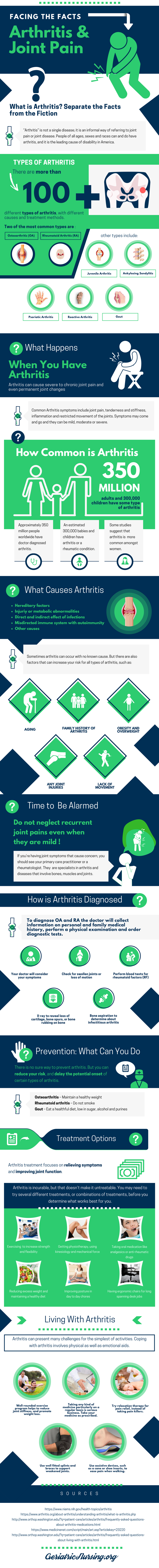
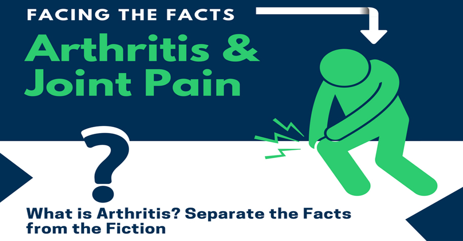
Arthritis- An infographic by GeriatricNursing.org
Website Sources for Arthritis
1. Understanding Arthritis. This is the site to start your search for arthritis answers. It’s a site created by the Arthritis Foundation and has a wealth of information on the site.
2. What is Rheumatoid Arthritis? This is a site designed by the Arthritis Foundation specifically about rheumatoid arthritis. It’s a good site for people who know they have RA and want to know the latest in diagnosis and treatment of this joint disease.
3. Let’s Dig Into Everything about RA-This is a site put out by a RA support organization that delves into rheumatoid arthritis and its management. They have resources for experimental RA treatment.
4. Septic Arthritis– This is a comprehensive review of septic arthritis designed for people who are health professionals or learned patients wanting to learn all they can about this condition.
5. Gout and Pseudogout– The patient with crystal arthritis will learn all they want to know on this comprehensive site. It’s designed for the person who wants to know the science and medicine behind these two types of arthritis.
6. The American College of Rheumatology puts out this information site for patients and caregivers who want to know about osteoarthritis and its manifestations.
7. Find a Rheumatologist – It isn’t always easy to find a rheumatologist near you if you have an arthritic condition. This site from the American College of Rheumatology will help you get the help you need from a board-certified rheumatologist.
8. Rheumatoid Arthritis – This is a medical site that shows pictures of patients with rheumatoid arthritis plus a comprehensive review of the pathophysiology, presentation, workup, and treatment of this type of arthritis.
9. Gout – This is a lecture series on gout that gives many slides showing pictures of gout and images that easily explain the disease state.
10. Osteoarthritis – This is a picture-filled slide presentation on osteoarthritis. For individuals wanting a visual image of what this disease looks like plus valuable information on the disorder, this is the site to visit.