Tag Archives: injury

Common Football Injuries

Common Football Injuries

CommonFootballInjuries_FBsize

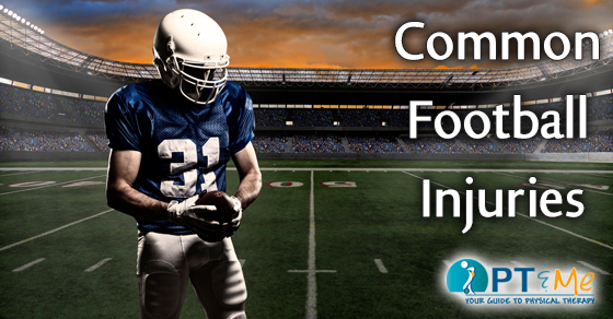
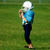
Football is one of the most popular sports played by young athletes, and it leads all other sports in the number of injuries sustained. In 2007, more than 920,000 athletes under the age of 18 were treated in emergency rooms, doctors’ offices, and clinics for football-related injuries, according to the U.S. Consumer Product Safety Commission. Physical therapy can provide specific treatment to a number of specific football injuries. Here are a few injuries that can happen during a football game or practice:

KNEE INJURIES
Knee injuries in football are the most common, especially those to the anterior or posterior cruciate ligament (ACL/PCL) and to the menisci (cartilage of the knee). These knee injuries can adversely affect a player’s long-term involvement in the sport. Football players also have a higher chance of ankle sprains due to the surfaces played on and cutting motions.

Physical therapy treatment for knee injuries may include:
• Exercises to help promote recovery. Specifically, therapists will design a program to strengthen the whole leg as well improve its range of motion.
• Balance exercises to allow the return to daily activities (including work and sports) while decreasing the risk of falls and reinjury
• Hands-on treatment to keep the knee joint from becoming stiff
• Ice and vasopneumatic pressure to reduce swelling and pain

SHOULDER INJURIES
Shoulder injuries are also common. The labrum (cartilage bumper surrounding the socket part of the shoulder) is particularly susceptible to injury, especially in offensive and defensive linemen. In addition, injuries to the acromioclavicular joint (ACJ) or shoulder are commonly seen in football players.

Physical therapy treatment for shoulder injuries may include:
NON-SURGICAL
Most labral tears will respond well to non-surgical treatment and may be just one component of a multi-factored pathology of the aging shoulder. Physical therapy will typically address a labral tear from the biomechanical approach of improving the motion and reducing the repetitive injury. If the inflammation and mechanical stress on the structures can be reduced then the tissue has a chance to heal.
SURGICAL
If the athlete has had surgery to the shoulder, the therapist will follow a specific protocol to apply just the right amount of strain on the shoulder to keep it safe after surgery. A sling may be recommended in the early stages but the therapist will get the arm moving with assistance within a relatively short period of time. Physical therapists will give instructions on how to provide varying levels of assistance to the arm for motion in safe planes in front of the body, and eventually throughout the entire range of motion. Once the tissues are healed, the therapist will begin to put resistance on the support structures in order to improve the mechanics of motion and reduce the risk of another injury.

kid football player

CONCUSSIONS
Football players are very susceptible to concussions. A concussion is a change in mental state due to a traumatic impact. Not all those who suffer a concussion will lose consciousness. Some signs that a concussion has been sustained are headache, dizziness, nausea, loss of balance, drowsiness, numbness/tingling, difficulty concentrating, and blurry vision. The athlete should return to play only when clearance is granted by a health care professional. It is recommended that players go though a concussion baseline test before the start of the season. Results from baseline tests (or pre-injury tests) can be used and compared to a similar exam conducted by a healthcare professional during the season if an athlete has a suspected concussion. More information here.

Physical therapy treatment for concussions may include:
EVALUATION: The physical therapist will take time to talk with you and perform a thorough examination of your condition.
THERAPY: The physical therapist will plan a treatment program suited to your individual condition, which will involve exercises for your balance, vision, inner ear and more in order to restore brain function.
TEACHING: Physical therapists will spend time reviewing information with you regarding your diagnosis and progress as well as answering your questions. This empowers the patient to make a lifelong impact on their health.
RETURN TO SPORT: Physical therapists are uniquely qualified to guide you towards a safe return to sport. A therapist can guide recovering athletes through a stepwise protocol to keep patients symptom free,  and to prevent serious, life-threatening conditions associated with a second head injury due to early return to football.

OVERUSE INJURIES
Low-back pain, or back pain in general, is a fairly common complaint in football players due to overuse. Overuse can also lead to overtraining syndrome, when a player trains beyond the ability for the body to recover.

Physical therapy treatment for overuse injuries may include:
Pain-relieving techniques (such as ice) and decreasing or modifying painful activities. This diagnosis often occurs from muscular tightness or weakness which causes posture to get out of alignment. A physical therapist will educate and assist in proper stretching and strengthening exercises for the back. They may perform hands on, manual therapy techniques to further increase joint flexibility. The final phase of rehab will involve strengthening during functional activities and education to prevent the injury from recurring.

RESOURCES:
U.S. Consumer Product Safety Commission
www.cpsc.gov

Stop Sports Injuries
www.stopsportsinjuries.org

REFERENCES:
Preventing Football Injuries. http://www.stopsportsinjuries.org/STOP/Prevent_Injuries/Football_Skating_Injury_Prevention.aspx

Backpack Safety

Backpack Safety 101

BackpackSafety_FBsize

It’s that time of the year again when the kids are heading back to school. Learn the proper guidelines and precautions about backpack safety in this week’s PTandMe blog post.

Size
• Should Not Extend Above Shoulders
• Should Rest In Contour Of Low Back (Not Sag Down Toward Buttocks)
• Should Sit Evenly In Middle Of Back

Fit
• Shoulder Straps Should Rest Comfortably On Shoulders And Underarms, With Arms Free To Move – Tighten Shoulder Straps To Achieve This Fit
• Tighten Hip And Waist Straps To Hold Pack Near Body
• Padded Straps Help Even Pressure Over The Shoulders

Weight Of Pack
• Should Never Exceed 15% Of The Child’s Weight To Avoid Excess Loads On The Spine

backpack

Lifting Of Pack
• Proper Lifting Is Done By Bending The Knees, Squatting To Pack Level, And Keeping Pack Close To Body To Lift First To Waist Level And Then Up To Shoulders

Carrying The Pack
• Keep Both Shoulder Straps In Place And Pack Centered
• Spinal Forces Increase With Distance From The Body’s Center

Posture
• Uneven Stresses On The Spine Can Cause Muscle Imbalances. This Can Lead To Pain And Possibly Functional Scoliosis.

Plantar Fasciitis

Hamstring Tightness and Plantar Fasciitis

Hamstring Tightness_FBsize

Plantar fasciitis is classically characterized by pain in the central to medial plantar heel. It is thought to be caused by chronic inflammation of the plantar fascia due to repetitive strain and trauma to the fascia. There are many other purported causes including, calcaneal spurs and increased intraosseous calcaneal pressure, among others. Many studies have shown inflammatory and histological changes at the origin of the plantar fascia and surrounding structures that are consistent with repetitive strain and degenerative changes including a thickening of the fascia.

Biomechanics has long been looked at when investigating possible causes of injuries. Alteration of the load-bearing characteristics of the foot has been suggested by several studies to be the underlying problem in plantar fasciitis. Microtears and chronic degenerative changes result from the increased tensile stress placed on the fascia due to the changes in biomechanics. Muscle tightness is one factor that can lead to changes in gait mechanics and load bearing of the foot. Hamstring tightness has recently been investigated as a factor in plantar fasciitis and has been shown to induce prolonged forefoot loading due to increased knee flexion during gait. A rapid progression through the contact phase of gait results from increased knee flexion and in turn increases forefoot pressure. The fascia is a fixed-length ligament, so an increase in forefoot pressure results in increased tension at its insertion on the calcaneus. The increased time spent on the forefoot in gait leads to a chronic traction injury that is localized to the hindfoot insertion of the fascia; which is consistent with the symptoms of heel pain.

plantar

Biomechanical deficits have long contributed to injuries. Only recently has hamstring tightness been shown to have an effect on plantar fasciitis. Hamstring tightness affects every step, resulting in a biomechanical deficit which may contribute to a tensile overload of the plantar fasciitis. Recent studies suggest that all patients with plantar fasciitis should be evaluated for hamstring tightness. Physical therapy treatment for plantar fasciitis should include hamstring stretching.

More information about Plantar Fasciitis can be found in the PTandMe injury center.

physical therapy near me

This information was written by Plymouth Physical Therapy Specialists. They are committed to using evidence-based treatments in their practice. This means that their therapists utilize the most current and clinically relevant treatments in their approach to rehabilitation. For more information click here.

Juvenile Rheumatoid Arthritis

Juvenile Rheumatoid Arthritis

Juvenile Rheumatoid Arthritis_FBsize

Definition

Juvenile rheumatoid arthritis (JRA), also known as juvenile idiopathic arthritis, is a disease of the joints in children. It can affect a child over a long period of time. JRA often starts before the child is 16 years old.

In Juvenile rheumatoid arthritis, the joint becomes swollen. It will make the joint painful and difficult to move. JRA can also lead to long term damage to the joint. For some, JRA can interfere with the child’s growth and development.

There are 5 major types of juvenile rheumatoid arthritis:
• Pauciarticular JRA—4 or less joints are affected in the first 6 months of illness
• Polyarticular JRA—5 or more joints are affected in the first 6 months of illness
• Enthesitis associated arthritis—swelling of the tendon at the bone
• Psoriatic arthritis—associated with a skin disease called psoriasis
• Systemic onset JRA (also called Stills disease)—affects the entire body, least common type of JRA

Causes

Juvenile rheumatoid arthritis is caused by a problem of the immune system. The normal job of the immune system is to find and destroy items that should not be in the body, like viruses. With JRA, the immune system attacks the healthy tissue in the joint. It is not clear why this happens. The immune system problems may be caused by genetics and/or factors in the environment.

People of Hispanic American, Asian American, Pacific Islander, Native American, or African American descent are at higher risk.
Having prediabetes means that you are at high risk for developing diabetes and may already be experiencing adverse effects of elevated blood sugar levels.

Risk Factors

Girls are more likely to get JRA than boys.

There are no clear risk factors for JRA. Factors that may be associated with some types of JRA include:
• Family history of:
• Anterior uveitis with eye pain
• Inflammatory back arthritis (ankylosing spondylitis)
Inflammatory bowel disease

kids toes

Symptoms

• Symptoms may include:
• Joint stiffness, especially in the morning or after periods of rest
• Pain, swelling, tenderness, or weakness in the joints
• Fever
• Weight loss
• Fatigue or irritability
• Swelling in the eye—especially associated with eye pain, redness, or sensitivity to light
• Swollen lymph nodes
• Growth problems, such as:
• Growth that is too fast or too slow in one joint (may cause one leg or arm to be longer than the other)
• Joints grow unevenly, off to one side
• Overall growth may be slowed

Some symptoms are specific to each type of juvenile rheumatoid arthritis . For example:
• Symptoms common with pauciarticular JRA include:
• Problems most often found in large joints. These joints include knees, ankles, wrists, and elbows.
• If the left-side joint is affected, then the right-side similar joint will not be affected. For example, if the right knee is affected, then the left knee will be healthy.
• May also have swelling and pain at on the tendons and ligaments attached to the bone

• Symptoms common with polyarticular JRA include:
• Problems found most often in small joints of the fingers and hands. May also affect weight-bearing joints like the knees, hips, ankles, and feet.
• Joints on both sides of the body are affected. For example, if the left hand is affected, then the right hand will also be affected.
• May also have a blood disorder called anemia. This is an abnormally low number of red blood cells.
• One type of polyarticular JRA may occur with:
• A low-grade fever
• Nodules—bumps on parts of body that receive a lot of pressure such as elbows

• Symptoms common with systemic onset JRA include:
• Some of the first signs may be a high fever, chills, and a rash on the thighs and chest. May appear on and off for weeks or months
• May have swelling in the heart, lungs, and surrounding tissues
• The lymph nodes, liver and/or spleen may become enlarged
• Children with enthesitis arthritis often have tenderness over the joint where the pelvis and spine meet.
• Children with psoriatic arthritis often have finger or toe swelling. There may also be damage on fingernails.

Often, there are remissions and flare-ups. Remission is a time when the symptoms improve or disappear. Flare-ups are times when symptoms become worse.

Diagnosis

You will be asked about your child’s symptoms. You will also be asked about your family medical history. A physical exam will be done. An eye examination may also be done to check for swelling in the eye. Your child may be referred to a specialist if JRA is suspected. The specialist is a doctor that focuses on diseases of the joints.

Images may be taken of your child’s bodily structures. This can be done with x-rays.

Your child’s bodily fluids may be tested. This can be done with:
• Blood tests
• Urine tests
• Tests of joint fluid

Treatment

Talk with your doctor about the best plan for your child. The plan will work to control swelling, relieve pain, and control joint damage. The goal is to keep a high level of physical and social function. This will help keep a good quality of life. Treatment options include the following:

Medication

There are several types of medication that may be used:

• Nonsteroidal anti-inflammatory drugs (NSAIDs)—to help swelling and pain
• Disease-modifying antirheumatic drugs (DMARDs)—to slow the progression of the disease
• Tumor necrosis factor (TNF) blockers—to decrease swelling, pain, and joint stiffness
• Interleukin inhibitors—to reduces disease activity
• Corticosteroids through IV or by mouth—for swelling
• Steroid injections into the joint—may help relieve swelling and pain in some children

Polyarticular JRA may become inactive in children who begin medications within 2 years of onset.

Physical Therapy

Exercise is done to strengthen muscles and to help manage pain. Strong nearby muscles will support the joint. It also helps to recover the range of motion of the joints. Normal daily activities are encouraged. Non-contact sports and recreational activities may be good options. Physical activities can also help boost a child’s confidence in their physical abilities.

Physical therapy may be needed. This will help to make the muscles strong and keep the joints moving well.

Maintenance Devices

Splints and other devices may be recommended. They may be worn to keep bone and joint growth normal. Some joints may get stuck in a bent position. These devices can help prevent this.

Prevention

There is no known way to prevent JRA.

by Jacquelyn Rudis

RESOURCES:
American College of Rheumatology
http://www.rheumatology.org

Arthritis Foundation
http://www.arthritis.org

CANADIAN RESOURCES:
The Arthritis Society
http://www.arthritis.ca

Health Canada
http://www.hc-sc.gc.ca

REFERENCES:

Hofer MF, Mouy R, et al. Juvenile idiopathic arthritides evaluated prospectively in a single center according to the Durban criteria. J Rheumatol. 2001. 28:1083.

Juvenile idiopathic arthritis (JIA) enthesitis related. EBSCO DynaMed website. Available at: http://www.ebscohost.com/dynamed. Updated September 16, 2015. Accessed December 21, 2015.

Juvenile idiopathic arthritis (JIA) oligoarticular. EBSCO DynaMed website. Available at: http://www.ebscohost.com/dynamed. Updated September 16, 2015. Accessed December 21, 2015.

Juvenile idiopathic arthritis (JIA) polyarticular. EBSCO DynaMed website. Available at: http://www.ebscohost.com/dynamed. Updated September 16, 2015. Accessed December 21, 2015.

Juvenile idiopathic arthritis (JIA) systemic-onset. EBSCO DynaMed website. Available at: http://www.ebscohost.com/dynamed. Updated September 16, 2015. Accessed December 21, 2015.

JAMA Patient Page. Juvenile idiopathic arthritis. JAMA. 2005;294:1722.

Petty RE, Southwood TR, et al. Revision of the proposed classification criteria for juvenile idiopathic arthritis: Durban, 1997. J Rheumatol.1998; 25:1991.

2/5/2013 DynaMed’s Systematic Literature Surveillance http://www.ebscohost.com/dynamed: De Benedetti F, Brunner HI, Ruperto N, et al. Randomized trial of tocilizumab in systemic juvenile idiopathic arthritis. N Eng J Med. 2012;367(25):2385-95.

2/24/2014 DynaMed’s Systematic Literature Surveillance http://www.ebscohost.com/dynamed: Limenis E, Grosbein HA, et al. The relationship between physical activity levels and pain in children with juvenile idiopathic arthritis. J Rheumatol. 2014 Feb;41(2):345-351.

9/2/2014 DynaMed’s Systematic Literature Surveillance http://www.ebscohost.com/dynamed: Guzman J, Oen K, et al. The outcomes of juvenile idiopathic arthritis in children managed with contemporary treatments: results from the ReACCh-Out cohort. Ann Rheum Dis. 2014 May 19.

Last reviewed December 2015 by Kari Kassir, MD Last Updated: 12/20/2014

EBSCO Information Services is fully accredited by URAC. URAC is an independent, nonprofit health care accrediting organization dedicated to promoting health care quality through accreditation, certification and commendation.

aquatic therapy

Is Aquatic Therapy For You?

Aquatic Therapy_FBsize

Aquatic therapy offers an alternative environment for therapeutic exercise. If you have tried traditional physical therapy, or have restrictions on your physical therapy program, aquatic Therapy may be the perfect solution for your physical therapy needs.

Why Aquatic Therapy is Beneficial

Less pressure: The buoyancy of the water decreases the amount of pressure, or compressive forces, on your joints and spine. When you’re immersed in water up to your neck, the weight pressing down on your body is reduced by 90%. When the water is up to your waist, the pressure is reduced by 50%.

Reduced swelling: The pressure of the water helps to move fluid from the injured area back into the body. Decreased swelling is essential for regaining the strength and motion needed for recovery.

Ease of movement:
Water is an element that supports and assists movement. It offers a safe setting for regaining strength and joint range of motion.

Faster progress: Aerobic conditioning can often be performed in the water, even when it may be too soon or too difficult to do in the clinic. Staying stable in the water, challenges your core and balance. Plus, sports specific activity can begin earlier than on land.

old man swimming

Who Can Benefit From Aquatic Therapy

• Chronic pain patients requiring a more gentle form of therapy
• Patients at risk of falls due to balance and gait disorders
• Patients with severe arthritis or other weight-bearing restrictions
• Prenatal and postnatal patients
• Patients with general deconditioning
• Sports medicine and orthopedic patients requiring an accelerated component to their rehab protocol

This information was written by Life Fitness Physical Therapy, a privately-owned, outpatient physical therapy practice operating 14 clinics in the metro and surrounding Baltimore, Maryland area. Life Fitness Physical Therapy was founded in 2000 based on the core values of providing the highest level of customer service and clinical excellence in outpatient physical therapy. For more information click here.

TMJ

There is Hope for TMJ Pain

HopeForTMJPain_FBsize

TMJ is where the lower jaw meets the skull.

The temporomandibular joint or TMJ, is a complex joint located in front of each ear. It is responsible for allowing mouth opening and closing. When the TMJ is not working correctly, you may experience jaw pain, clicking, popping, or locking of the joint. You may also experience headaches, neck pain, sinus pain, dizziness, and ear ringing or pain. TMJ pain or Temporomandibular Dysfunction (TMD) is not strictly limited to the jaw, it can also be influenced by the alignment of the head and neck.

The goal of Physical Therapy treatment for Temporomandibular Dysfunction (TMD) is to provide pain relief using a variety of techniques to improve your range of motion through exercise and manual therapy and to improve posture and muscle imbalance. There may be modifications to your activities or work station that would be helpful as well.

man with ear pain

SYMPTOMS
• Face or Jaw Pain
• Ear Pain
• Tinnitis (ringing in the ears)
• Dizziness
• Headache
• Jaw Clicking and/or Popping
• Neck Pain
• Limited Jaw Opening

TREATMENT
Your Physical Therapist will work closely with your Dentist or Physician to establish an individualized treatment plan based on results from a comprehensive evaluation. The most current treatment options will be utilized to ensure the best outcome for decreased pain and improved function.

TREATMENT WILL INCLUDE, BUT IS NOT LIMITED TO:
• Comprehensive evaluation of head, neck, and jaw
• Education regarding the diagnosis and related head, neck and jaw dysfunction. This includes addressing any of your questions or concerns.
• Individualized program specific to your needs including modalities, joint mobilization, soft tissue mobilization, postural education, neuromuscular re-education and TMJ management techniques.
• Collaboration between referring physician or dentist to ensure comprehensive and integrated care.

causes of pitching arm injuries

The Most Common Causes of Pitching Arm Injuries

PitchingArmInjuries_FBsize

The most common causes of pitching arm injuries to high school, college and professional pitchers are overload, overuse, lack of proper conditioning and improper throwing mechanics.

• Overload is the result of throwing too many pitches during one outing. Maximum pitch counts for various age groups, or for an individual pitcher’s normal strength and stamina, are effective in preventing overload.

• Overuse is the result of pitching too often and not having an adequate recovery time or a good arm maintenance program (stretching, running, strengthening, throwing) between pitching assignments. Coaches and trainers should be aware that each individual pitcher varies in arm strength, arm fatigue, arm tightness and soreness and require different recovery time needs. Make certain you provide your pitcher with a good active recovery program between pitching assignments.

• Proper conditioning involves the entire body; the legs and core muscles as well as the throwing arm. Coaches should supervise a proper stretching and warm-up procedure that is performed daily before throwing a ball.

• Pre-season is one of the most frequent times for arm injuries. Pitchers throw too much and too hard, too early. Also, they have not ingrained their normal rhythm and often are attempting to learn new techniques or new pitches. Another major factor of injury during pre-season is that pitchers are not working with a normal in-season rotation schedule and do not get enough recovery time from a lot of necessary drill work that involves throwing (pick-offs, defensive plays, etc.).

• Improper throwing mechanics. If a pitcher has improper throwing techniques, with the body or arm, there is a great chance of early fatigue of the throwing mechanism, and of course injury. The more power and force generated, the greater the chance of injury. In a competitive situation, most pitchers will not admit they are fatigued, experiencing minor pain or have a minor injury. It is important that you, a coach, trainer or physical therapist be able to recognize changes in the pitcher’s mechanics, performance, or mannerisms.

night pitcher

This information about common causes of pitching arm injuries was written by University Physical Therapy, an outpatient physical therapy group with eight locations in New River Valley, Virginia. University PT is THE choice for outstanding sports rehabilitation, physical therapy and occupational therapy services. For more information click here.

What Should I Tell My Physical Therapist?

What_Should_I_Ask_FBsize

If you have never been to a physical therapist before you may have some questions about your care and expectations. Here are some general questions and points about physical therapy you may want to talk about during your initial evaluation.

Talk about the pain you are experiencing.
Go into detail about what sort of activities you have trouble with and where the pain is coming from.  The more details you can provide the more it will help your therapist develop a treatment plan.

How did your injury happen and/or when did your pain begin?
Did you have an injury or accident, or did the pain develop gradually over an extended period of time? Whether it’s sports, work or surgical post-op related your PT can guide you to recovery following an injury. As part of physical therapy, they can teach you exercises, stretches, and techniques using specialized equipment to address your pain.

What are your recovery goals?
If you have specific goals in relation to your recovery, let your therapist know. Do you want to be able to run a marathon, or is the goal to cook dinner and be able to reach up into your cabinets? Maybe your goal is to have enough strength to play with the grand kids. Your Physical Therapist needs this information when designing a treatment plan that best suits your needs. Whether you’re a professional athlete who’s suffered an injury or an orthopedic patient who needs assistance following surgery, your recovery starts with physical therapy.

old_guy_at_PT

Give your physical therapist you primary care provider’s information.
Good communication between your physical therapist and your primary care provider can help eliminate the need for unnecessary drugs/medications and provide great detail about your medical history. Your physical therapist can help clarify referral guidelines and the decision-making process as well.

Tell your physical therapist about any medications you are currently taking.
Different medications can cause adverse side effects due to physical activity and your physical therapist wants to make sure you are ready for physical therapy. Make sure you have a detailed list of your medications and be ready to discuss them at your evaluation.

Always tell your physical therapist your pain levels and how you feel your progress is going.
Physical Therapy is intended to make changes in your body.  As physical therapists make changes to your tissues and mechanics, the stresses on your body will change, and may change where you feel the pain or symptoms. Be open with your physical therapist. If you have any questions about your treatment or want to go over any pain you are experiencing let your physical therapist know and they can go over it with you. Chances are what you are experiencing is completely normal and part of the healing process.

For more information about physical therapy programs and treatment techniques visit our About Physical Therapy section by clicking here.

Need to find a physical therapist? Visit our Find A PT Page

removing kinesio tape

Removing Your Kinesio Tex Tape

RemovingKinesio_FBsize

Removing Kinesio Tape can seem pretty daunting. The whole point is for it to stay on, so what do you do when it’s time to remove it? Fear not friends, we have the answers here.

DON’T TAKE IT OFF TOO FAST! It’s not a Band-Aid.

1. Put some baby oil or cooking oil on the tape and let it soak for a few minutes to break down the adhesive.
2. Remove the tape in the direction of hair growth by slowly rolling down the tape and applying pressure on the skin with your other hand.
3. Use oil to remove any extra residue.

kinesio

TIP: The tape can be worn for 3-5 days as long as there is no skin irritation.

To learn more about Orthopedic Kinesio taping visit our treatment technique section here

This information about removing Kinesio Tape was written by Desert Hand and Physical Therapy, a physical therapy group with locations throughout the greater Phoenix and Mesa AZ area. Desert Hand and Physical Therapy has specialized treatment centers providing therapeutic treatment and rehabilitation to restore optimal upper extremity function. For more information about the group click here.

More PTandMe articles about Kinesio Taping can be found here:

kinesio taping pain relief   kinesiotaping helpful for lymphedema patients

PT News

This Month in PT News. Featuring articles from PTandMe partnering clinics!

GymBike

1. The Rapid Recovery Blueprint
Written by the Therapy Team at Cornerstone Physical Therapy

Exercise tends to break down muscle fibers while rest allows muscles to recuperate and emerge stronger. The appropriate amount of exercise, followed by recovery leads to an ongoing cycle of improvement over time. Strength and endurance improve, creating a foundation for long-term health. Read more

GymSeniors

2. Can We Get Stronger as We Age?
Written by the Therapy Team at the Jackson Clinics Physical Therapy

The answer to that question is – absolutely! After age 40 or so, we all begin to lose muscle strength and bone density, and our hormone production slows. Read more

shoulderInjury

3. Overtraining Doesn’t Help, It Hurts
Written by the Therapy Team at Momentum Physical Therapy

Working to achieve a sports or fitness goal can drive many people to overtrain in an effort to get stronger, better, faster. Our PT’s have seen many injuries related to overtraining. Read more